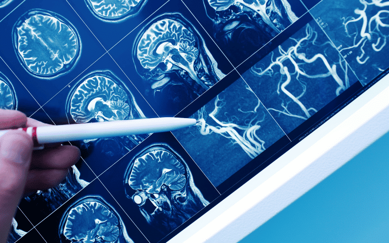
Fact 6: The Crucial Role of Imaging Tests in Cerebrovascular Disease

Imaging tests play a vital role in diagnosing cerebrovascular disease. These tests give doctors a detailed look at the structures within the brain and can help identify any abnormalities. Let’s take a closer look at some of these tests.
Computed tomography (CT) is often the first test used to evaluate a person with suspected cerebrovascular disease. A CT scan can quickly identify areas of the brain that have been damaged by stroke and can also detect bleeding.
Magnetic resonance imaging (MRI), on the other hand, uses a magnetic field and radio waves to create detailed images of the brain. An MRI can identify smaller or more subtle areas of brain damage than a CT scan.
An angiogram, which involves injecting a dye into the blood vessels to make them visible on an X-ray, can be used to view the blood vessels in the brain. This test can help identify blockages, aneurysms, or other abnormalities.
Transcranial Doppler ultrasound is another method used to monitor blood flow in the arteries of the brain. It can be particularly useful in assessing the risk of stroke in individuals with sickle cell disease. (6)