7. Through the Lens: The Role of Brain Scans in Diagnosing Pick’s Disease

Brain imaging techniques are invaluable tools in the field of neuroscience, allowing a visual exploration of the brain’s landscape. In the context of Pick’s disease, these scans provide key insights into the structural changes within the brain, thus assisting in its diagnosis and differentiation from other dementia forms.
Imagine for a moment being able to take a journey inside the brain, witnessing firsthand the intricate network of neurons and synapses. In a healthy brain, it’s akin to navigating a vibrant city, bustling with activity. However, in Pick’s disease, certain parts of this city – particularly the frontal and temporal lobes – begin to shrink and lose their vitality. This process is called ‘atrophy’, a term commonly encountered in the realm of neurodegenerative diseases.
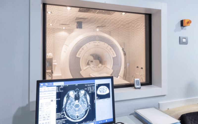
Magnetic Resonance Imaging (MRI) scans can create detailed images of the brain, shedding light on this atrophy. The MRI acts like a high-definition camera, capturing the intricate details of the brain’s structure. In Pick’s disease, it reveals the areas where significant shrinkage has occurred, providing clues to the underlying pathology.
Similarly, Computed Tomography (CT) scans provide another perspective, offering a different kind of visual representation. Like comparing a cityscape viewed from a helicopter versus on foot, each gives unique insights into the structural changes in the brain. CT scans can highlight abnormalities in brain structure and, in conjunction with an MRI, can offer a more comprehensive picture of the disease’s impact.
While these imaging techniques do not confirm the diagnosis of Pick’s disease on their own, they form an integral part of the diagnostic process. The patterns of atrophy they reveal, when combined with clinical symptoms and history, can provide a robust framework for diagnosing Pick’s disease accurately. (7)