7. Radiographic Imaging: The Key to Accurate Diagnosis
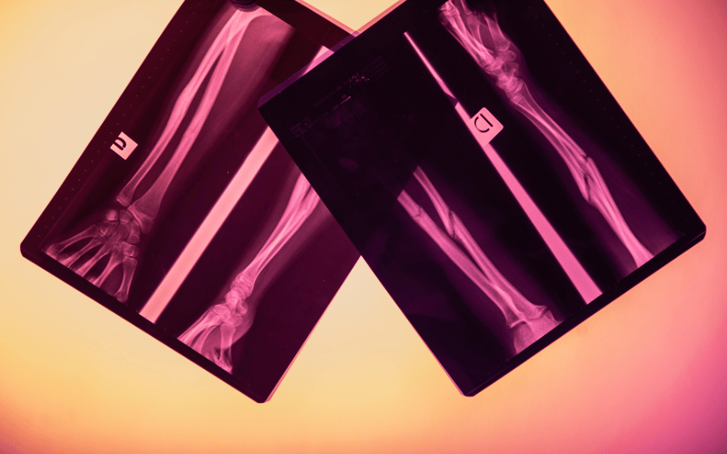
Radiographic imaging is indispensable in the diagnosis of greenstick fractures. Given the often subtle nature of these fractures, X-rays are typically the first line of investigation. They provide a clear image of the bone, revealing the characteristic partial break or bend indicative of a greenstick fracture. Advanced imaging techniques, such as MRI or CT scans, are rarely necessary but can be employed in complex cases where more detailed visualization is needed.
The role of radiographic imaging extends beyond mere identification. It aids in determining the exact location and extent of the fracture, which is critical in planning the appropriate course of treatment. The images can reveal whether the fracture is displaced or involves a growth plate, information crucial for predicting healing outcomes and potential complications.
Interestingly, the development of digital radiography has significantly enhanced the diagnostic process. Digital X-rays offer higher image quality and allow for easier manipulation and analysis, leading to more precise diagnoses. They also expose patients to less radiation, an important consideration in pediatric care.
Radiographic imaging in the context of greenstick fractures also demonstrates the technological advancements in medical diagnostics. These tools not only provide clarity in diagnosis but also contribute to a better understanding of pediatric bone injuries. They exemplify the marriage of technology and medicine in improving patient outcomes, particularly in the nuanced field of pediatric orthopedics. (7)
