4. The Role of Medical Imaging in Diagnosis

Diagnosing Subglottic Stenosis is a complex process, requiring a combination of clinical evaluation and advanced imaging techniques. Medical imaging plays a pivotal role in this, providing detailed views of the airway that are crucial for accurate diagnosis and effective treatment planning.
Computed Tomography (CT) scans are a commonly used tool in the diagnosis of Subglottic Stenosis. They provide cross-sectional images of the airway, allowing medical professionals to assess the extent of the narrowing and any associated abnormalities.
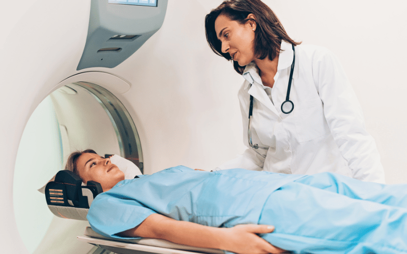
Magnetic Resonance Imaging (MRI) offers another layer of detail, providing high-resolution images of both the airway and surrounding tissues. This is particularly useful in cases where the extent of the condition is unclear or where additional information is needed to guide treatment.
In some cases, direct visualization of the airway through a procedure known as bronchoscopy may be necessary. This procedure involves inserting a small camera through the nose or mouth, providing a real-time view of the airway and any obstructions.
These imaging techniques, combined with a thorough clinical evaluation, ensure that individuals receive the most accurate diagnosis possible. This, in turn, lays the groundwork for effective, tailored treatment, improving outcomes and quality of life for those affected by Subglottic Stenosis. (4)