7. Diagnostic Puzzles: Deciphering KS
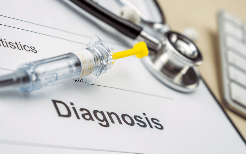
Diagnosing Kaposi Sarcoma isn’t always straightforward. Its lesions, while distinct, can be mistaken for other skin conditions. Moreover, with the possibility of internal tumors, the diagnostic process often extends beyond mere visual assessments.
For many clinicians, a skin biopsy becomes the tool of choice. A small sample, when observed under the microscope, can reveal the presence of the spindle-shaped tumor cells typical of KS. But this is just one piece of the puzzle. The presence of HHV-8 in the biopsy further consolidates the diagnosis.
In cases where internal lesions are suspected, endoscopy becomes a crucial tool. Whether it’s the lungs or the digestive tract, specialized scopes provide a glimpse into the body’s recesses, hunting for those elusive KS tumors.
Medical imaging, from X-rays to MRIs, can further aid the quest, especially when the disease is believed to have infiltrated the deeper organs. Each image, each shadow, becomes a clue, guiding the clinician towards a definitive diagnosis. (7)
