Fact 13: The Role of Neuroimaging

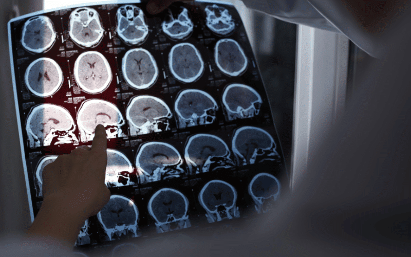
The field of neuroimaging has made tremendous strides over the years, significantly benefiting the understanding and diagnosis of cognitive impairment and dementia. Neuroimaging techniques give us the ability to look directly at the brain in ways we could never do before. It provides a glimpse into the physical and functional changes that accompany these conditions, contributing valuable information to the diagnostic process.
Structural imaging techniques, such as computed tomography (CT) and magnetic resonance imaging (MRI), can provide detailed images of the brain’s anatomy. These scans can reveal physical changes like atrophy (shrinkage of brain tissue), strokes, or other damages to the brain tissue that could explain cognitive symptoms. For example, in Alzheimer’s disease, an MRI might reveal significant shrinkage in the hippocampus, a brain region critical for memory.
Functional imaging techniques, including positron emission tomography (PET) and functional magnetic resonance imaging (fMRI), offer a different perspective. They allow clinicians to assess the brain’s activity levels and metabolic processes. For example, in Alzheimer’s disease, a PET scan might show reduced glucose metabolism in specific brain regions, suggesting those areas aren’t functioning as they should be. (13)