Introduction: A Closer Examination on Pyogenic Granuloma
Pyogenic granuloma, also known as lobular capillary hemangioma, presents a unique challenge in dermatology. This benign vascular lesion, often mistaken for other skin conditions, has perplexed both patients and medical professionals alike. In this article, we delve into the intricacies of pyogenic granuloma, shedding light on its characteristics, causes, and effective treatment strategies.

First and foremost, it’s crucial to recognize that pyogenic granuloma is more than just a cosmetic concern. The condition, characterized by small, red, and often bleeding bumps on the skin, can have a significant impact on an individual’s quality of life. These lesions, although non-cancerous, can be alarming due to their sudden appearance and tendency to bleed easily.
Understanding what triggers pyogenic granuloma is key to managing it effectively. While the exact cause remains unknown, factors like trauma, hormonal changes, and certain medications have been linked to its development. This correlation highlights the importance of a thorough medical history and examination in diagnosing the condition.
The journey to an accurate diagnosis can be fraught with confusion. Pyogenic granuloma mimics several other skin conditions, making it essential for healthcare professionals to conduct a comprehensive assessment. This often includes a biopsy, ensuring that the treatment plan addresses the specific needs of the condition.
In summary, pyogenic granuloma is a condition that demands attention and understanding. By exploring its causes, symptoms, and treatment options, we aim to provide valuable insights for those affected by it. Stay tuned as we unfold the layers of pyogenic granuloma, offering guidance and clarity on this perplexing skin condition.
1. The Nature of Pyogenic Granuloma: A Closer Look at Its Appearance

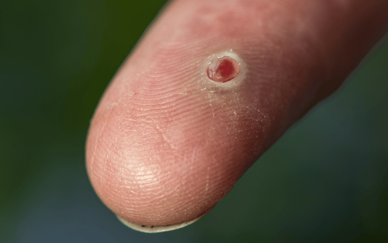
Pyogenic granuloma, a benign vascular lesion, often presents itself in a distinctive manner. The appearance of these lesions is a crucial aspect in understanding this condition. Typically, pyogenic granulomas manifest as small, red, and tender bumps on the skin. They are known for their rapid growth, often developing over a few weeks.
The color of these lesions can range from pink to red, sometimes with a brownish hue, indicating a high concentration of blood vessels. The surface of a pyogenic granuloma is usually smooth but can become rough and crusted if it bleeds, which is a common occurrence. These growths are most often found on the hands, arms, and face, but they can appear anywhere on the body.
Interestingly, the size of pyogenic granulomas can vary significantly. They typically measure a few millimeters in diameter, but some can grow to several centimeters. Despite their alarming appearance, these lesions are not cancerous. However, due to their propensity for bleeding and ulceration, they can cause discomfort and concern.
The texture of pyogenic granulomas adds to their distinctive profile. They are generally soft to the touch and can feel like a small balloon filled with fluid. This texture is due to the lesion’s composition, primarily of blood vessels and inflammatory cells.
In summary, the appearance of pyogenic granulomas is a telltale sign of this condition. Their distinctive color, size, and texture make them recognizable to those familiar with dermatological conditions. Understanding these characteristics is key to identifying pyogenic granuloma and differentiating it from other skin lesions. (1)