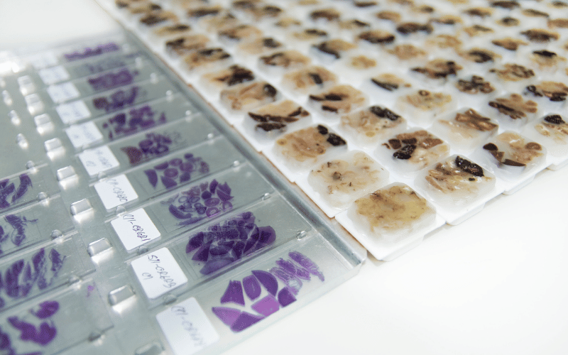
11. The Histopathological Perspective: Understanding Granuloma at the Cellular Level

The study of pyogenic granuloma at the cellular level, known as histopathology, reveals intricate details about this condition. Understanding its histopathological aspects is key to grasping how these lesions develop and behave.
Under the microscope, pyogenic granuloma presents a characteristic appearance. It consists of a proliferative growth of capillaries, small blood vessels, in a lobular arrangement, hence the term ‘lobular capillary hemangioma.’ This growth is usually accompanied by a mix of inflammatory cells, indicating the body’s response to the lesion.
The capillaries in a pyogenic granuloma are often surrounded by a fibromyxoid stroma, a type of connective tissue. This tissue provides a matrix for the capillaries and contributes to the lesion’s overall structure. The presence of numerous immature and proliferating capillaries is a hallmark of this condition, distinguishing it from other vascular lesions.
The histopathological examination also aids in differentiating pyogenic granuloma from malignant conditions. Despite its rapid growth and sometimes alarming appearance, the lack of atypical cells and controlled growth pattern signify its benign nature.
In summary, the histopathological examination of pyogenic granuloma provides a deeper understanding of its nature. This microscopic perspective is not only crucial for accurate diagnosis but also enriches our comprehension of the condition’s development and behavior. (11)