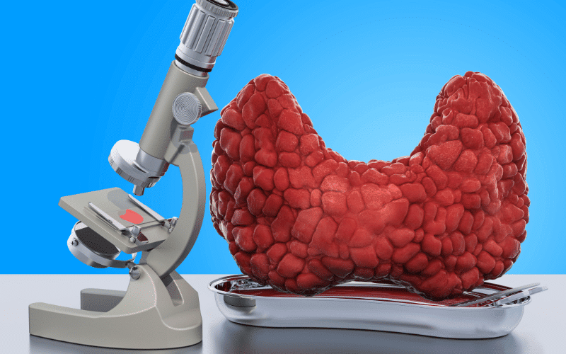
3. Histopathological Features: PTC vs. FTC – Diving into the Microscopic World

The crux of differentiating between Papillary and Follicular Thyroid Cancer often lies in their microscopic features, providing us valuable insights into their nature, behavior, and potential treatment strategies.
PTC, true to its name, is characterized by the presence of papillae—branching structures reminiscent of tiny trees, when viewed under a microscope. These papillae are lined by cells exhibiting distinctive nuclear features such as clearing, overlapping, and grooves, earning them the term “Orphan Annie Eye” nuclei. Another common characteristic of PTC is the formation of psammoma bodies, tiny calcium deposits that can be identified in the tumor tissue.
On the flip side, FTC paints a different histological picture. It is characterized by a predominance of follicles, small spherical structures lined by thyroid cells. Importantly, the distinction between benign follicular adenomas and FTC relies on one key feature: capsular and/or vascular invasion. This means that to confirm an FTC diagnosis, there must be evidence of the tumor breaching the capsule surrounding the thyroid or invading the blood vessels.
Furthermore, the nuclei of FTC cells are generally less distinct and do not exhibit the same “Orphan Annie Eye” appearance as PTC. Psammoma bodies, too, are typically absent in FTC.
Therefore, the microscopic features offer a clear demarcation between PTC and FTC, reinforcing their unique identities. As we delve deeper into their structural intricacies, we realize the integral role that these histopathological characteristics play in the clinical management of thyroid cancers. (3)