Fact 3: Diagnostic Difficulties of Small Vessel Disease
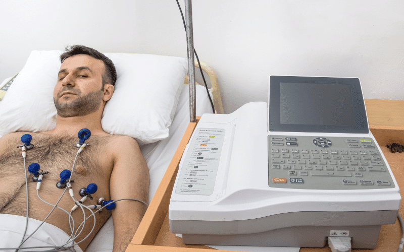
Diagnosing small vessel disease presents its own unique challenges. A condition as elusive as SVD often slips through the cracks of conventional diagnostic tools. An electrocardiogram (ECG) is a prime example of this issue. ECGs are a staple in detecting heart-related problems, yet they fail to accurately portray the state of the smallest arteries affected by SVD.
The ECG’s shortcoming stems from its nature. It monitors the heart’s electrical activity, making it an excellent tool for identifying arrhythmias and ischemic heart disease. However, it is not designed to visualize or assess the condition of the arteries. Thus, the effects of small vessel disease, especially in the early stages, often remain undetected during ECG examinations.
In recent years, coronary angiograms have been used to detect SVD. This invasive procedure involves injecting dye into the coronary arteries and taking X-ray images. The dye helps to visualize the blood vessels and can indicate areas of blockage or narrowing. However, the technique is not foolproof, especially in the case of SVD where the changes occur in the very small arteries. (3)
