Fact 4: The Discovery – X-Ray Vision into Dental Health
The revelation of an odontoma’s presence is often serendipitous, a byproduct of routine dental imaging. It’s within the grayscale nuances of an X-ray that the odontoma’s signature becomes apparent. These growths proclaim their existence in shadows and contrasts, emerging on dental films as areas of increased density amidst the landscape of the jawbone.
The discovery of an odontoma is a testament to the value of diagnostic imaging in dentistry. It’s not an overstatement to say that X-rays provide a superpower of sorts—a vision that transcends flesh and bone, unveiling the secrets hidden within the dental anatomy. For odontomas, which are frequently asymptomatic, this vision is crucial in bringing them to the attention of dental professionals.
The radiographic appearance of odontomas is characteristic. Compound odontomas manifest as a cluster of tiny, tooth-like structures, whereas complex odontomas present as a chaotic radiopacity without a discernible pattern. This distinction is critical for the ensuing treatment plan, as the type, size, and location gleaned from the X-ray inform the surgical approach.
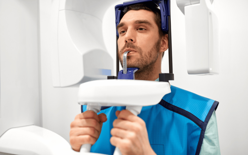
Continued advancements in dental imaging, including 3D cone-beam computed tomography (CBCT), further enhance the discovery process. These technologies allow for a three-dimensional assessment, providing unparalleled detail and aiding in the precise localization of the odontoma, which is essential for minimally invasive surgery.
In summary, the narrative of odontoma discovery is a tale of shadows unveiled. It underscores the profound impact that radiographic technology has on dental diagnostics, ensuring that even the silent whispers of abnormal growths do not go unnoticed in the pursuit of dental well-being. (4)
