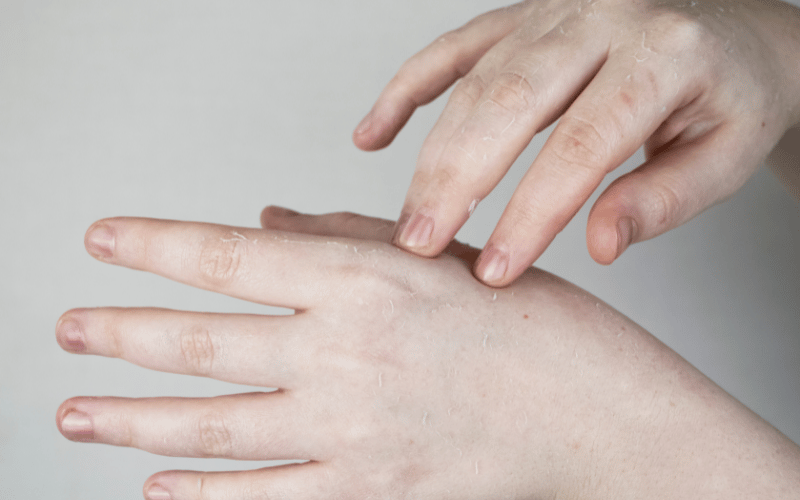
2. Plaques: The Raised Red Flags of Mycosis Fungoides

As the disease progresses, the patches often evolve into thicker lesions known as plaques. These are raised, often reddish-brown areas that resemble a more severe dermatological issue.
The texture of these plaques can vary. Some might feel rough to the touch, while others might have a smoother, shinier finish. They’re not just “upgraded” patches. Plaques signal a deeper involvement of the skin layers by the malignant T-cells and underscore the importance of addressing the condition promptly.
Plaques form due to the proliferation and accumulation of abnormal T-cells in the skin’s deeper layers. This “build-up” pushes the skin outward, leading to the raised appearance.
Moreover, as these cells continue to grow uncontrollably, they interfere with the skin’s natural processes, often leading to inflammation and further discoloration. This cellular activity differentiates the plaques of mycosis fungoides from other skin conditions.
While plaques can technically form anywhere on the body, they have a tendency to appear in certain regions more than others. The folds of the skin, such as the armpits, groin, and areas under the breasts, are particularly susceptible. This pattern is likely due to the higher moisture content and skin friction in these areas, which provides a conducive environment for the malignant T-cells to thrive.
Identifying plaques early and seeking intervention is vital. The transition from patches to plaques indicates an advancement in the disease. (2)